For more information on the management of these tumors *** click here***. You will link to Dr. Ojemann's web page on meningiomas.
.
GLIOMA
These primary brain tumors originate in brain tissue. Metastatic
brain tumors spread to the brain tissue through the blood stream predominately. Gliomas
are often designated as benign or malignant types although further subtypes are generally
determined after biopsy tissue is evaluated by the neuropathologist. The treatments
vary based on the clinical condition of the patient and the growth potential of the
tumor type. The effects of the tumor vary based on the area of the brain involved
and can also include seizures or headaches or symptoms of increased intracranial
pressure from the mass effect of the tumor and surrounding cerebral edema. MRI scans
generally clearly defne the tumor and the area of the brain involved and can be used
to follow the growth of the tumor and side effects of edema or hydrocephalus. Surgery
is often performed to remove or at least biopsy the tumor using stereotactic techniques
to precisely localize the tumor within the skull. Radiation therapy or chemotherapy
are often utilized in the treatment of the malignant gliomas. Surgical implantation
of chemotherapy wafers directly on the tumor surface can be considered especially
for recurrent malignant glioblastoma multiforme tumors. Tumor protocols have also
included treatment by infecting the cerebral gliomas first with a virus and then
following surgery, giving anti-viral drugs The tumor mass can be reduced by the effects
of drugs on infected cells and the inflammatory response of the brain against the
infected tissue. Gamma Knife stereotactic radiation can be used as a primary treatment
if the diagnosis is already determined or for small recurrent tumor re-growth. The
gold standard is usually surgical resection of all tumor microscopically followed
by radiation therapy. Decadron is often given for 3 days or more pre-operatively
to reduce cerebral edema. All the options required careful clinical individual evaluation
by your physician and discussion of the treatment alternatives.
Basic Information on
Gliomas
Images and description
of a Malignant Glioma
Information on
Stereotactic Biopsy
MENINGIOMA
(read below)
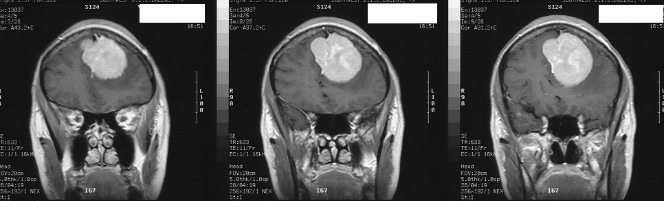
THESE TUMORS ORIGINATE FROM THE MENINGES OR COVERING OF THE BRAIN OR SPINAL CORD.
MENINGIOMAS CAN BECOME QUITE LARGE BEFORE SYMPTOMS AND SIGNS OF BRAIN OR SPINAL CORD
DYSFUNCTION ARE NOTICED BY THE PATIENT. OFTEN SUBTLE PROBLEMS WITH CEREBRAL FUNCTION
ARE DISCOVERED SUCH AS PERSONALITY CHANGES OR MEMORY IMPAIRMENT OR LONG STANDING
HEADACHES WHEN I QUESTION THE PATIENT. OCCASIONALLY, SMALL MENINGIOMAS CAN BE ASSOCIATED
WITH SURROUNDING CEREBRAL EDEMA OR CAUSE SEIZURES. TUMORS THIS LARGE NEED TO BE REMOVED
BY CAREFUL MICROSURGERY AND OUTCOMES CAN BE EXCELLENT AS IN THIS CASE.
For more
information on the management of these tumors *** click
here***. You will link to Dr. Ojemann's web page on meningiomas.
.
Updated 10-26-98