
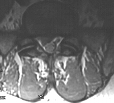
A Herniated Disc at the L4-L5 level can be seen clearly on the MRI below. The
disc is named L4-L5 because of the location between the 4th Lumbar and the 5th Lumbar
Vertebral bodies.
Importantly, the clinical condition of the patients is critical
in deciding how to best treat this spinal disorder. Many individuals improve with
passage of time, avoidance of strenuous activities such as bending and lifting and
bed rest. Often additional appropriate conservative medical treatment such as non-steroidal
anti-inflammatory medications and physical therapy exercises will be needed. Questions
need to be asked and a neurological examination performed in order to assess a patient's
clinical condition. Follow up evaluation to determine the results of treatment will
be important in making a decision on additional medical treatment or surgery. The
decision to perform a Lumbar Microscopic Discectomy is based on many factors. For
instance, is this painful condition acute or chronic and is the patient's leg function
impaired or is the pain incapacitating? Does the patient have progressive neurological
deficits or severe neurological impairment of a lumbar nerve root? Does the patient
have impairment of bladder function such as hesitancy, incontinence or genital/rectal
region numbness? What diagnostic tests have been done and does the patient's neurological
condition correlate directly with the MRI or CT scan or myelogram images? Are there
other medical conditions present or problems co-existing at a seperate level of the
spine? Discussion with the patient regarding the outcome of surgery and possible
complications and the expected benefit is extremely important. Long term care for
their spinal condition has to be considered.
The sagittal MRI image reveals a large disc herniation at L4-L5 with extruded fragments extending above the disc space within the spinal canal. The lower axial image shows a disc herniation with compression of the nerve roots within the spinal canal at L2-L3 on a different individual.
Updated 8-12-98