LUMBAR SYNOVIAL CYSTS
Synovial cysts are most frequently discovered nowadays
with MRI scanning. Previous studies such as myelograms and post-myelogram CT scans
were quite useful especially when viewing the skeletal or bony anatomy of the facet
joints. These lesions can be present for long periods of time on the surface of
the lumbar facet joints. Synovial cysts have also been found to have formed rather
quickly over months instead of years on repeat MRI scans of the lumbar spine. Osteoarthritis
affecting facet joints or history of trauma or repetitive traumatic strain on the
joints or spinal motion segments are often discovered during physician evaluation.
Synovial
cysts can enlarge and create pressure on the nerve roots within the spinal canal
or foramen under the facet joints. Leg pain often radicular in quality, numbness,
weakness or bladder control problems can occur with nerve root compression in the
spinal canal. Local back pain is often associated with these lesions. Microsurgical
findings include firm cheesy masses often with small amount of fluid, non-infectious,
extruded from the medial aspect of the facet joint. The expansion into the spinal
canal often causes inflammation and significant adherence of this mass to the nerve
root or dura. Cerebral spinal fluid leakage can occur even with meticulous microsurgical
dissection when removing the lesion. Often thickened ligamentum flavum or elastic
yellow ligament between the lamina and inside of the facet is found in conjunction
with the synovial cyst.
The results of surgery are often dramatic with improvement
of nerve root function and reduction in pain. Long term care includes avoidance
of increasing weight gain and reconditioning exercises to strengthen the spinal
musculature. Walking exercises, aquatics exercises, bicycle use often are comfortably
performed in moderation. Corsets to support the spine can be useful but not as beneficial
as strong supporting musculature and proper posture along with proper biomechanical
lifting techniques.
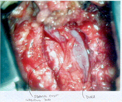
Left sided white-yellow cyst pushing the purple-blue dura
to the right. Inside the dura are the cauda equina nerve rootlets and under the
mass was the L5 traversing nerve root at this L4-5 level of the lumbar spine.
